


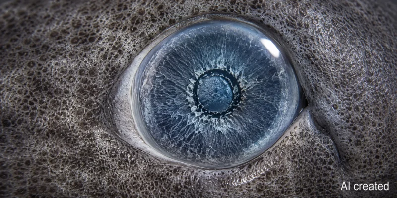
Greenland sharks were long considered nearly blind due to their dark Arctic environment, small eyes, and the copepods frequently found on their corneas. A study in Nature Communications paints a different picture: even in animals over a century old, the retina, light-sensitive cells, and the molecular machinery for low-light vision remained intact.
A report published on August 22, 2026, by ScienceDaily revisits these findings. However, the research paper itself is not new; it was originally published on January 5, 2026. This distinction is important for context, as the current headline describes not a second dataset, but a new round of media coverage regarding the same study.
What the team examined in the eyes
The researchers combined histology, genome and RNA analyses, fluorescent probes for various retinal cells, lipid measurements, and functional tests of the visual pigment rhodopsin and the cornea. The samples came from Greenland sharks caught using scientific longlines off Disko Island, Greenland, between 2020 and 2024.
The methods complement one another but rely in part on very small sample sizes. Histological and several cellular analyses were performed on the retina of a single animal; lipid measurements were conducted on two retinas, and RNA analysis on three individuals. To assess light transmission, the team examined six preserved corneas. The results therefore provide strong evidence of function at the tissue and molecular levels, though they do not yet offer a complete description of vision in the wild.
A retina designed for low light
The retina examined contained densely packed, elongated rod cells but no cones. Rods are particularly sensitive to even a few photons, whereas cones—in many vertebrates—enable vision in bright light and color discrimination. Consistent with this, the genes required for rod vision were intact and active, while numerous genes associated with the cone system were missing, had become non-functional pseudogenes, or were no longer being expressed.
The visual pigment is also adapted to the depth at which the shark lives. Its rhodopsin absorbed light most strongly at 458 nanometers—within the blue spectrum. Short-wavelength blue light penetrates particularly far in clear water and dominates the faint residual light of the Arctic deep sea. Consequently, the Greenland shark likely does not see fine details or colors the way a diurnal animal does, but it possesses a highly specialized system for detecting faint light signals.
Eye parasites do not automatically mean blindness
Many Greenland sharks carry the copepod *Ommatokoita elongata* on their corneas. All six corneas examined showed signs of parasitism at their edges. Nevertheless, they allowed 70 to 100 percent of light to pass through across the measured range; in the blue spectrum of 450 to 500 nanometers, transmission was between 66 and 100 percent. This does not rule out the possibility that the parasites cause localized damage or impair vision. However, it demonstrates that a parasitized cornea is not necessarily opaque.
More than 130 years old – without detectable retinal degeneration
The oldest animal examined was estimated to be over 130 years old. Nevertheless, all retinal layers were present, the cell types required for rod-mediated vision were detectable, and no obvious signs of cell death or DNA fragmentation were observed in the sections examined. This finding applies to the samples in question; it does not prove that every very old Greenland shark is free from all forms of age-related vision deterioration.
The study highlights DNA repair as a possible explanation. Long-lived shark species retained the gene ercc1, and ercc4 was expressed more strongly in the retina of the Greenland shark than in the species used for comparison. Both belong to the repair complex ERCC1-XPF. This is a promising lead, but not yet proof that this mechanism alone protects the retina over decades; further functional experiments are required to establish this.
What “not blind” means in scientific terms
The study does not measure visual acuity, the field of view, or hunting behavior. Instead, it demonstrates that light can reach the cornea, the retina remains structurally intact, key cell types are present, and genes associated with rod-based vision remain active. Together, these findings provide compelling evidence of a functional visual system adapted to darkness—though not necessarily one capable of sharp vision under all conditions.
Potential applications in human medicine are also in their infancy. Gene activity in an extremely long-lived cold-water shark cannot be directly translated into treatments for macular degeneration or glaucoma. Its primary value lies in providing a natural model that allows researchers to study the long-term preservation of nerve tissue.
Another piece of the puzzle regarding extraordinary longevity
This new analysis complements two other lines of research we have previously covered: estimates of age and maturity reveal just how slowly the species grows and how late it reaches reproductive age. An examination of their highly aged hearts revealed clear signs of aging but no demonstrable loss of function. The eyes and heart do not tell the same story here; yet, together, they demonstrate that extreme longevity in this species does not simply equate to a uniform decline of all organs.
For conservation efforts, the implications are ambiguous: while the retention of vision over such a long period is a remarkable adaptation, it does not render the species any less vulnerable. Slow growth, very late sexual maturity, and bycatch in Arctic fisheries mean that the loss of adult individuals can be replenished only extremely slowly.
