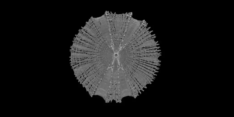
How a shark swims appears to be written deep inside its vertebrae. Florida Atlantic University reports on a study in the Journal of Anatomy, in which a research team compared the three-dimensional architecture of the spines of six shark species with very different swimming styles. The study was published online on July 14, 2026, followed by the detailed research release on July 27.
The findings challenge an oversimplified view of the shark skeleton. Although the spines of cartilaginous fishes are not made of bone, their vertebral centra contain mineralized plates called lamellae and branching nodes. Their number, shape and spatial arrangement vary among species and change from the front of the body toward the tail.
A three-dimensional comparison of 139 vertebrae
The team examined 139 vertebrae from 24 sharks. Where available, samples came from anterior, middle and posterior sections of the spine. High-resolution micro-computed tomography revealed the mineralized structures without destroying the vertebrae during imaging.
The researchers then measured the size and shape of the centra, the volume of mineralized tissue, and the lamellae and branches. This allowed them to compare not only which species have strongly mineralized vertebrae, but also how the internal blueprint changes along the body.
Stiff power transfer in fast sharks
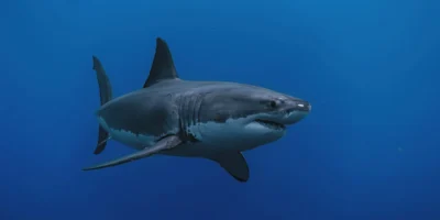
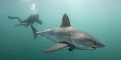
The fast open-ocean swimmers Great White Shark (Carcharodon carcharias), Shortfin mako (Isurus oxyrinchus) and Porbeagle (Lamna nasus) shared a similar basic design. Their largest vertebrae occurred in the middle of the body. Toward the tail the vertebrae became smaller but contained more lamellae. This combination may stabilize the mid-body and stiffen the posterior spine so that muscular power is efficiently transferred to the tail fin.
That pattern fits the thunniform swimming style of these species: much of the body remains comparatively still, while rapid lateral movement is concentrated around the caudal peduncle and tail. The study therefore shows that speed is reflected not only in muscles and external body shape, but also in the internal architecture of the vertebrae.
The thresher shark is the extreme case
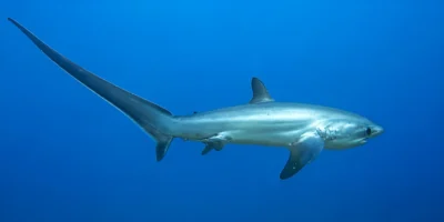
Common thresher shark (Alopias vulpinus) was especially striking. Its vertebrae contained the highest number of mineralized lamellae and branches. At the same time, the posterior vertebrae were strongly compressed lengthwise and provided a large contact area with adjacent vertebrae.
The researchers interpret this combination as an adaptation to exceptional loads. Common thresher sharks not only accelerate their long upper tail lobe, but also strike schooling fish from the side or over the body while hunting. Their spine must withstand forces from several directions, remain stable and still allow the mobility required for a tail strike.
Some of the thresher shark material had already been examined in a 2024 study by the same team. The new work places those vertebrae in a broader comparison of six species from four families of mackerel sharks.
Maneuverability instead of maximum speed
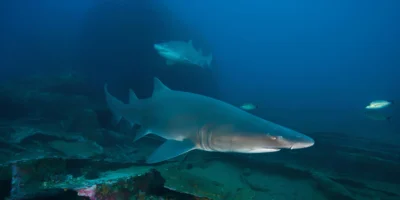
The researchers found a different emphasis in Sand tiger shark (Carcharias taurus). Large anterior vertebrae became smaller along the spine, lamella counts were lower and branching was more extensive. The team interprets this pattern as a more flexible spine suited to slow, maneuverable swimming in structurally complex habitats.
Basking shark (Cetorhinus maximus) represented the opposite extreme from the fast species. Its long, deeply concave vertebrae were only weakly mineralized. This fits a slow-swimming filter feeder that experiences high drag while feeding with its mouth open, but does not need a stiff spine for explosive acceleration.
A blueprint with limitations
The functional explanations are well-supported biomechanical hypotheses, not direct load tests. The animals were not measured while swimming, and the individual vertebrae were not pushed to their mechanical limits under controlled forces. The researchers infer function from the three-dimensional structures, known swimming styles and regional patterns along the spine.
Despite that limitation, a shark spine is clearly not a uniform rod of cartilage. Evolution has placed mineralized material where a species needs stiffness, stability or mobility. The same principle could inspire lightweight technical materials that must be both strong and flexible, including applications in robotics and medical technology.